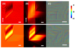
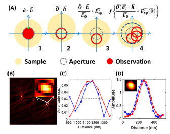
Facilitated by nanofabrication and optical analysis techniques, the development of plasmonics has seen extensive applications in optical trapping, imaging, and biosensing. Based on interferometric plasmonic microscopy, an optical approach capable of imaging single exosomes in a label-free manner has been proposed, which contributes to clinical exosome analysis and further understanding of exosome–antibody binding kinetics. A method for direct control of Ca2+ channels, referred to as femtosecond laser activated store-operated calcium channel (femtoSOC), has been developed based on ultrafast laser without the need of optogenetic tools or any other exogenous reagents, hence alleviating the requirements of optogenetic activation of SOCs. A system to obtain quantitative amplitude and phase images in interferometric plasmonic microscopy has also been developed, which can be applied to bionanoparticle analysis and characterizing nanoplasmonic devices.


Quantitative amplitude and phase imaging with interferometric plasmonic microscopy
Selected Publications:
1. Yang Y, Shen G, Wang H, et al. Interferometric plasmonic imaging and detection of single exosomes. Proceedings of the National Academy of Sciences, 2018, 115(41): 10275-10280.
2. Cheng P, Tian X, Tang W, et al. Direct control of store-operated calcium channels by ultrafast laser. Cell Research, 2021, 31: 758–772.
3. Yang Y, Zhai C, Zeng Q, et al. Quantitative amplitude and phase imaging with interferometric plasmonic microscopy. ACS Nano, 2019, 13: 13595-13601.
4. Jiang X, Pu R, Wang C, et al. Noninvasive and early diagnosis of acquired brain injury using fluorescence imaging in the NIR-II window. Biomedical Optics Express, 2021, 12(11): 6984-6994.
5. Wang S, Liu Y, Zhang D, et al. Photoactivation of extracellular-signal-regulated kinase signaling in target cells by femtosecond laser. Laser & Photonics Reviews, 2018, 12: 1700137.
